Asbestos Exposure: Who Is At Risk?


Asbestos was one of the most common industrial materials put to use in the twentieth century. It has also proven to be one of the most lethal, as inhaling asbestos fibers can lead to a wide range of pulmonary problems such as asthma and asbestosis - and can also be the direct cause of mesothelioma.

What is mesothelioma? It is a lethal cancer that attacks the membranes around the lungs, the heart and the abdominal cavity. Mesothelioma cancer of the lungs is by far the most common form. Perhaps its most unusual characteristic is that mesothelioma diagnosis usually occurs decades after the initial exposure to asbestos.

It takes years for the asbestos fibers to work their way into those membranes; after an extended presence they begin to cause fluid accumulation and tumor development. However the first mesothelioma symptoms are such afflictions as a persistent cough or shortness of breath - symptoms that are often mistaken for evidence of more common lung problems, which delays the mesothelioma diagnosis even further.

Los Angeles Personal Injury Attorneys


Brain Injury
Over the course of a lifetime, the costs of a traumatic brain injury (TBI) can be tremendous. Depending of the severity of the injury, a person who has suffered a traumatic brain injury may be permanently unable to care for him or herself. Even in more moderate injury cases, a person who has suffered any brain damage may have difficulty remembering basic things and suffer from dizzy spells.

If you or someone you love has suffered a traumatic brain injury due to someone else's negligence, come to the law firm of Lederer & Nojima LLP in Los Angeles, California. Contact us today to schedule your free initial consultation.
Working with a Team of Experts to Protect You

When you come to our firm, we'll prepare your legal claims to address the difficulties you will face for the rest of your life. In traumatic brain injury cases we routinely work with experts, including life care planners, who can help to fully assess the lifetime costs of such a serious injury. We also rely upon many of the top professionals in Southern California to provide expert medical testimony for our personal injury cases.

Traumatic brain injuries can arise in a wide range of circumstances. A car accident might result in permanent brain damage. A construction accident may result in permanent brain injuries and spine injuries. The mechanics of the various injuries may differ but the effect is identialc: i.e. the loss of function and capacity due to brain damage.
Personal Attention and Convenient Representation for Personal Injury Claims

We provide personal attention for all of our personal injury cases. When you come to the law firm of Lederer & Nojima LLP, you will always deal directly with a personal injury attorney - not a paralegal or legal secretary. We will provide you with updates regarding your case throughout our legal representation of your claims.

We understand that it is often difficult for seriously injured people to travel. If you are homebound or in the hospital, we can come to visit you. We handle all of our personal injury cases on a contingency fee basis - you will pay no attorneys fees until we recover compensation for you.

Contact us today to schedule your free consultation. We often successfully represent people who have been turned down by other attorneys. Call us at 310-694-5907 to discuss your case.

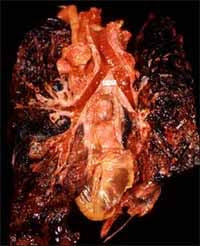
What Is pericardial mesothelioma?


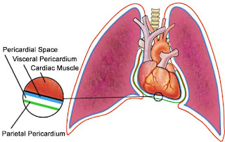
Pericardial mesothelioma cancer is found in less than 10 percent of mesothelioma patients. While pleural mesothelioma is fairly well understood (inhaled asbestos fibers become imbedded in the lining of the lung and lead to cancerous tumors),….. pericardial mesothelioma is not. Pleural mesothelioma develops because the inhaled asbestos fibers are very durable and cannot be eliminated through the body’s processes. Over time, these fibers cause chronic inflammation that eventually leads to growth of cancerous tumors or, in some cases, asbestos. In contrast, researchers and doctors are unsure how the asbestos fibers become lodged in the heart’s serous lining, or pericardium. One theory holds that asbestos fibers are broken into smaller pieces in the lungs and then carried into the blood stream. From there, proponents of this theory hypothesize, the fibers are pumped through the heart, become lodged in the heart lining, and cause the chronic inflammation that leads to cancerous growths (as with pleural mesothelioma).

For pericardial mesothelioma, it is not clearly understood how the asbestos fibers become lodged in the heart serous lining or pericardium. It is possible that asbestos fibers, after being broken into smaller pieces in the lungs, are carried from the lungs into the blood stream. As the fibers are pumped through the heart, they become lodged in the heart lining. Once lodged in the pericardium, the chronic inflammation process leading to cancerous growths is similar to that occurring in pleural mesothelioma.

Because pericardial mesothelioma is so rare, there is little specific research on this type of mesothelioma. Approximately 2,000 new cases of mesothelioma are diagnosed each year, and fewer than 200 of them are pericardial mesothelioma.

Symptoms of Pericardial Mesothelioma


As with both the pleura and the peritoneum, the growth of tumors in the pericardial region causes the expansion of tissue and allows fluid to accumulate around the heart The fluid can interfere with the functioning of the heart……. It is this fluid that is responsible for most of the symptoms of pericardial mesothelioma. These symptoms include:

Chest pain, ranging from uncomfortable to severe
Shortness of breath
Heart palpitations
Persistent coughing
Extreme fatigue after minimal activity or exertion
Pericardial mesothelioma resembles the other types of asbestos-caused mesothelioma in that the symptoms are often mistaken for something more common. In this case, heart attack or some other sort of heart disease is suspected, especially when the patient begins complaining of persistent chest pains.

Symptoms can vary with each patient and can be affected by a number of variables, including the location and size of the tumors and the overall health of the patient. The age of the patient may also factor into the equation.

A thorough patient history is the key to the proper diagnosis of the disease. Anyone who worked with asbestos or asbestos-containing products, particularly in shipyards or in the construction field, should always mention their exposure to their doctor. Because this is a rare disease, other diagnoses are usually considered before doctors investigate the possibilities of mesothelioma.

What Causes Pericardial Mesothelioma?


Prolonged exposure to asbestos is - to this day - the only known cause of pericardial mesothelioma as well as the other types of the disease. With mesothelioma, loose asbestos particles are inhaled and gather in or near the internal organs, most often the pleural, which is the lining of the lungs…..

There is no exact answer as to how an individual develops pericardial mesothelioma and how the dangerous asbestos fibers make their way into the pericardium. Researchers theorize that asbestos fibers, once they are broken up into smaller pieces in the lungs, are carried from the lungs into the blood stream. As the fibers are pumped through the heart, they may become lodged in the heart lining. Once lodged in the pericardium, the chronic inflammation process, which leads to the formation of cancerous growths or tumors, is similar to what occurs in the more common pleural mesothelioma.

What is Lung Cancer?


Cancer of the lung, like all cancers, results from an abnormality in the body’s basic unit of life, the cell. Normally, the body maintains a system of checks and balances on cell growth …..so that cells divide to produce new cells only when needed. Disruption of this system of checks and balances on cell growth results in an uncontrolled division and proliferation of cells that eventually forms a mass known as a tumor.

Tumors can be benign or malignant; when we speak of “cancer,” we refer to those tumors that are considered malignant. Benign tumors can usually be removed and do not spread to other parts of the body. Malignant tumors, on the other hand, grow aggressively and invade other tissues of the body, allowing entry of tumor cells into the bloodstream or lymphatic system and then to other sites in the body. This process of spread is termed metastasis; the areas of tumor growth at these distant sites are called metastases. Since lung cancer tends to spread or metastasize very early in its course, it is a very life-threatening cancer and one of the most difficult cancers to treat. While lung cancer can spread to any organ in the body, certain organs — particularly the adrenal glands, liver, brain, and bone — are the most common sites for lung-cancer metastasis.

The lung is also a very common site for metastasis from tumors in other parts of the body. Tumor metastases are made up of the same type of cells as the original, or primary, tumor. For example, if prostate cancer spreads via the bloodstream to the lungs, it is metastatic prostate cancer in the lung and is not lung cancer.

Lung Cancer:Signs and Symptoms


People often decide to visit the doctor only after they have been bothered by certain complaints over a period of time. Individuals who have lung cancer frequently experience symptoms such as the following:

* Cough
* Shortness of breath
* Wheezing
* Chest pain
* Hemoptysis (bloody, coughed-up sputum)
* Loss of appetite
* Weight loss
* Pneumonia (inflammation of the lungs)

her symptoms that are associated with lung cancer include:

* Weakness
* Chills
* Swallowing difficulties
* Speech difficulties or changes (e.g., hoarseness)
* Finger/nail abnormalities (e.g., “clubbing,” or overgrowth of the fingertip tissue)
* Skin paleness or bluish discoloration
* Muscle contractions or atrophy (shrinkage)
* Joint pain or swelling
* Facial swelling or paralysis
* Eyelid drooping
* Bone pain/tenderness
* Breast development in men

Lung Cancer,Causes


It is estimated that tobacco smoking causes 80% of lung cancer deaths in men and 75% of lung cancer deaths in women. The more cigarettes that are smoked each day and the younger the age at which smoking began, the greater the risk of lung cancer. Lung cancer death rates are lower among individuals who use filter cigarettes and low-tar/low-nicotine brands, but such rates are significantly increased when compared with death rates in nonsmokers.


Advertising Disclaimer
People who quit smoking have a lower risk of developing lung cancer, and, after 10 to 15 years, their lung cancer death rates approach the rates of nonsmokers. Pipe and cigar smokers have higher lung cancer death rates than nonsmokers, but they have lower lung cancer death rates than cigarette smokers.

"Competing risk factors" such as secondhand smoke, asbestos, radon, occupational exposures, age, race, sex, and heredity also may play a role in lung cancer development. Additionally, there are some studies suggesting that lung cancer may be prevented by dietary micronutrients such as carotenoids, vitamin C, vitamin E, and selenium.

Smoking
Cigarette smoking is distinctly the greatest risk factor for lung cancer. There is a significant "dose-response" relationship between the number of pack-years smoked and lung cancer risk; that is, the more a person smokes and the longer he or she smokes, the greater the risk of lung cancer.

How exactly does cigarette smoking cause lung cancer? This question has not yet been answered definitively. But the most likely cause is the toxic mix of chemicals found in tobacco smoke. The major cigarette smoke chemicals that have been studied with respect to lung cancer are polycyclic aromatic hydrocarbons (PAHs), nicotine, nicotine by-products, tobacco-specific nitrosamines (TSNAs), metals such as nickel and cadmium, and radioactive polonium 210 (210Po).


About Tobacco . . .

Types of Tobacco In the United States, the main commercially grown species of tobacco is Nicotiana tabacum. Within this species, the types of tobacco used in cigarette products for America, western Europe, and Japan include bright (Virginia, flue-cured), Burley, Maryland, and oriental (aromatic) tobaccos. Cigarettes made in the United Kingdom and Finland primarily use bright tobaccos. Both varieties of cigarettes, when ignited, create smoke particles containing nicotine. Such "mainstream" smoke particles—minus water and nicotine—are the "tar" produced by burning cigarettes.

Tobacco Additives Cigarettes are made from sheets of reconstituted tobacco. Special solutions are used to keep the tobacco mixture intact, and chemicals known as humectants are added to maintain tobacco moisture. In addition, trade secret flavorants are included to make the product taste better during smoking. Such tobacco additives, when burned, may yield undesirable compounds. For example, the burning of licorice flavorant may produce chemicals known as polycyclic aromatic hydrocarbons (PAHs), and the burning of sugar may increase tar and nicotine in the smoke. Coumarin, a known cancer-causing substance in animals, has in the past been used as a tobacco additive. It is also likely that commercial tobaccos contain up to a few parts per million of the pesticides DDT and DDD, as well as the agricultural chemical maleic hydrazide.


About 400 to 500 separate gaseous substances are present in the smoke of a nonfilter cigarette. The major elements of cigarette vapor include nitrogen, oxygen, carbon dioxide, carbon monoxide, and water; other noteworthy substances include nitrogen oxides, hydrogen cyanide, formaldehyde, benzene, and toluene. The particles of cigarette smoke contain at least 3500 individual compounds such as nicotine, tobacco alkaloids (nornicotine, anatabine, anabasine), polycyclic aromatic hydrocarbons (PAHs; e.g., benzo(a)pyrene, B(a)P), naphthalenes, aromatic amines, phenols, and tobacco-specific nitrosamines (TSNAs).


Advertising Disclaimer
Tobacco-specific nitrosamines (TSNAs) are formed during tobacco curing and processing. TSNAs are chemicals that are suspected of causing lung cancer in humans. In rodent studies, regardless of the where or how it is applied, the TSNA known as NNK produces lung adenomas -benign tumors of epithelial (surface cell) tissue—and lung adenocarcinomas—malignant epithelial tumors with gland-like characteristics. The TSNA known as NNAL also produces lung adenocarcinomas in rodents, although it is a more powerful pancreatic carcinogen (cancer-causing substance) in rats.

Some of the TSNAs found in cigarette smoke particles are N-nitrosonornicotine (NNN), 4-(N-methyl-N-nitrosamino)-1-(3-pyridyl)-1-butanone (NNK), and N-nitrosoanatabine (NAB). Other TSNAs include 4-(N-methyl-N-nitrosamino)-1-(3-pyridyl)-1-butanol (NNAL), N-nitrosoanatabine (NAT), N-nitrosoanabasine (NAB), 4-(N-methyl-N-nitrosamino)-4-(3-pyridyl)-1-butanol (iso-NNAL), and 4-(N-methyl-N-nitrosamino)-4-(3-pyridyl)butyric acid (iso-NNAC). The U.S. National Academy of Science estimates that a pack-a-day smoker is exposed to about 17 micrograms (mg) of cancer-causing TSNAs.

Cigarette smoke contains the following approximate TSNA levels:


Approximate TSNA Levels in Cigarette Smoke

NNN NAT NAB NNK Total TSMAs
Nonfilter 278 236 30 156 700
Filter 209 172 21 156 558


Manufacturers can reduce the levels of TSNA in cigarette smoke by using lighter tobacco blends and by selecting parts of the tobacco plant that are low in nitrate—a forerunner of TSNAs. Yet tobacco blends with low amounts of nitrate may have higher levels of polycyclic aromatic hydrocarbons (PAHs) in mainstream smoke.

Polycyclic aromatic hydrocarbons (PAHs) also are believed to be major contributors to lung cancer risk in smokers. PAHs are "procarcinogens" that are metabolized, or broken down by the body into reactive substances. For example, the chemical benzo(a)pyrene is changed into a compound that is known to react with human genetic material (DNA) and form DNA "adducts." It is thought that such adducts may cause problems with lung cell reproduction that eventually may lead to lung cancer. Lung cancer patients who still smoke have higher levels of PAH-DNA adducts than smokers without lung cancer.

Table 2 provides estimates of PAH levels in cigarette smoke.


PAH levels in Cigarette Smoke (ng/cigarette)
Benzo(a)pyrene 10–50
5-Methylchrysene 0.6
Dibenz(a,h)anthracene 40
Benzo(b)fluoranthene 30
Benzo(j)fluoranthene 60
Chrysene 40–60
Benzo(e)pyrene 5-40


The International Agency for Research on Cancer (IARC) has declared that some PAHs in tobacco smoke show "sufficient" evidence of cancer-causing effects (carcinogenicity) in laboratory animals. Such PAHs include benzo(a)pyrene, benz(a)anthracene, benzo(b)fluoranthene, benzo(j)fluoranthene, benzo(h)fluoranthene, chrysene, dibenz(a,h)anthracene, dibenzo(a,i)pyrene, dibenzo(a,l)pyrene, indeno(1,2,3-cd)pyrene, and 5-methylchrysene.

Other chemicals within tobacco may damage the lungs. Tobacco contains at least 30 metals, although the most toxic of these—nickel and cadmium—are present in only small quantities. Most metals found in tobacco come from the soil, fertilizers, or agricultural sprays. The element polonium 210 (210Po), which is a radioactive compound, also has been identified in the particulate portion of cigarette smoke (0.03 - 0.07 pCi per cigarette).

Lung Cancer,Exams and Tests


Upon hearing about the symptoms, a health-care provider will formulate a list of possible diagnoses. He or she will ask questions about the symptoms, medical and surgical history, smoking and work history, and other questions about lifestyle, overall health, and medications.

Unless severe hemoptysis is occurring, a chest x-ray will most likely be performed first to look for a cause of the respiratory symptoms.

The x-ray film may or may not show an abnormality.


Types of abnormalities seen in lung cancer include a small nodule or nodules or a large mass.


Not all abnormalities observed on a chest x-ray are cancers. For example, some people develop scarring and calcium deposits in their lungs that may look like tumors on a chest x-ray film.
In most cases, a CT scan or MRI of the chest will further define the problem.

If symptoms are severe, the x-ray may be skipped and a CT scan or MRI may be performed right away.


The advantages of CT scan and MRI are that they show much greater detail than x-rays and are able to show the lungs in three dimensions.


These tests help determine the stage of the cancer by showing the size of the tumor or tumors.


They can also help identify spread of the cancer into nearby lymph nodes or certain other organs.
If a person's chest x-ray film or scan suggests that a tumor is present, he or she will undergo a procedure for diagnosis.

This procedure involves collection of sputum, removal of a small piece of the tumor tissue (biopsy) or a small volume of fluid from the sac around the lung.


The retrieved cells are reviewed under a microscope by a doctor who specializes in diagnosing diseases by looking at cell and tissue types (a pathologist).


Several different ways exist to obtain these cells.
Sputum testing: This is a simple test that is sometimes performed to detect cancer in the lungs.

Sputum is thick mucus that may be produced during a cough.


Cells in the sputum can be examined to see if they are cancerous. This is called cytologic review.


This is not a completely reliable test. If negative, the findings usually need to be confirmed by further testing.
Bronchoscopy: This is an endoscopic test, meaning that a thin, flexible, lighted tube with a tiny camera on the end is used to view organs inside the body.

Bronchoscopy is endoscopy of the lungs. The bronchoscope is inserted through the mouth or nose and down the windpipe. From there, the tube can be inserted into the airways (bronchi) of the lungs.


A tiny camera transmits images back to a video monitor.


The physician operating the bronchoscope can look for tumors and collect samples of any suspected tumors.


Bronchoscopy can usually be used to determine the extent of the tumor.


The procedure is uncomfortable. A local anesthetic is administered to the mouth and throat as well as sedation to make bronchoscopy tolerable.


Bronchoscopy has some risks and requires a specialist proficient in performing the procedure.
Needle biopsy: If a tumor is on the periphery of the lung, it usually cannot be seen with bronchoscopy. Instead, a biopsy is taken through a needle inserted through the chest wall and into the tumor.

Typically, a chest x-ray or CT scanning is used to guide the needle.


This procedure is safe and effective in obtaining sufficient tissue for diagnosis. After the chest surface is cleaned and prepared, the skin and the chest wall are numbed.


The most serious risk with this procedure is that the needle puncture may cause an air leak from the lung (pneumothorax). This air leak occurs in as many as 3%-5% of cases. Although this condition can be dangerous, it is almost always recognized and treated without serious consequences.
Thoracentesis: Lung cancers, both primary and metastatic, can cause fluid to collect in the sac surrounding the lung. This fluid is called a pleural effusion.

The fluid usually contains cells from the cancer.


Sampling this fluid can confirm the presence of cancer in the lungs.


The fluid sample is removed by a needle in a procedure similar to needle biopsy.


Thoracentesis can be important for both staging and diagnosis of the condition.
Thoracotomy: Sometimes a lung cancer tumor cannot be reached by bronchoscopy or needle procedures.
In these cases, the only way to obtain a biopsy is by performing an operation.


The chest is opened (thoracotomy), and as much of the tumor as possible is removed surgically. The removed tumor is then examined microscopically.


Unfortunately, this operation may not be successful in removing all tumor cells if the tumor is large or has spread to the lymph nodes outside of the lungs.


Thoracotomy is a major operation that is performed in a hospital.
Mediastinoscopy: This is another endoscopic procedure. It is performed to determine the extent that the cancer has spread into the area of the chest between the lungs (the mediastinum).

A small incision is made into the lower part of the neck, above the breastbone (sternum). A variation is to make the incision in the chest.


A mediastinoscope is inserted behind the breastbone.


Samples of the lymph nodes are taken to evaluate for cancer cells.


Mediastinoscopy is a very important step to determine whether the tumor can be surgically removed or not.
Other tests: Other tests are performed to stage the tumor and to assess a person's ability to withstand surgery and other treatment.

Pulmonary function tests assess breathing capacity.


Blood tests are performed to identify any chemical imbalances, blood disorders, or other problems that might complicate treatment.


CT scans or MRIs may be performed on the most common areas of spread to check for metastatic disease. These tests are generally performed only if symptoms occur that suggest metastatic disease. Certain treatment protocols require that these tests be performed.


A bone scan can determine whether the cancer has spread to the bones.
Staging: Staging is a method of classifying the tumor for purposes of treatment planning.

Staging is based on size of the tumor, location of the tumor, and degree of metastasis of the tumor (if any).


The treatment will be individually tailored to the tumor stage.


Tumor stage is related to the outlook for cure and survival (prognosis). The higher the tumor stage, the less likely the disease will be cured.
Lung cancer,Chemotherapy


In the original sense, a chemical that binds to and specifically kills microbes or tumor cells. The term chemotherapy was coined in this regard by Paul Ehrlich (1854-1915).
2. In oncology, drug therapy for cancer. Also called "chemo" for short.

Most cancer chemotherapeutic drugs are given IV (into a vein) or IM (into muscle). Some anticancer agents are taken orally (by mouth). Chemotherapy is usually systemic treatment, meaning that the drugs flow through the bloodstream to nearly every part of the body.

Patients who need many rounds of IV chemotherapy may receive the drugs through a catheter (a thin flexible tube). One end of the catheter is placed in a large vein in the chest. The other end is outside the body or attached to a small device just under the skin. Anticancer drugs are given through the catheter.

Chemotherapy is generally given in cycles: a treatment period is followed by a recovery period, then another treatment period, and so on. Usually a patient has chemotherapy as an outpatient at the hospital, at a doctor's office or clinic, or at home. However, depending on which drugs are given and the patient's general health, the patient may need to stay in the hospital for a short time.

The side effects of chemotherapy depend mainly on the drugs and the doses the patient receives. Most anticancer drugs affect cells that divide rapidly. These include blood cells, which fight infection, help the blood to clot, or carry oxygen to all parts of the body. When blood cells are affected by anticancer drugs, patients are more likely to develop infections, may bruise or bleed easily, and may have less energy. Cells that line the digestive tract also divide rapidly. As a result of chemotherapy, patients can have side effects, such as loss of appetite, nausea and vomiting, hair loss, or mouth sores. For some patients, medicines can be prescribed to help with side effects, especially with nausea and vomiting. These side effects tend to gradually go away during the recovery period or after treatment stops.

Hair loss, another side effect of chemotherapy, is a major concern for many patients. Some chemotherapy drugs only cause the hair to thin out, while others may result in the loss of all body hair. Patients may feel better if they decide how to handle hair loss before starting treatment.

In some men and women, chemotherapy drugs cause changes that may result in a loss of fertility (the ability to have children). Loss of fertility can be temporary or permanent depending on the drugs used and the patient's age. For men, sperm banking before treatment may be a choice. Women's menstrual periods may stop and they may have hot flashes and vaginal dryness. Periods are more likely to return in young women.

In some cases, bone marrow transplantation and peripheral stem cell support are used to replace blood cell production when it has been destroyed by chemotherapy and/or radiation therapy.

Lung cancer,Radiation therapy


The use of high-energy rays to damage cancer cells, stopping them from growing and dividing. Like surgery, radiation therapy is a local treatment that affects cancer cells only in the treated area.

Radiation can come from a machine (external radiation or from an a small container of radioactive material implanted directly into or near the tumor (internal radiation). Some patients receive both kinds of radiation therapy. External radiation therapy is usually given on an outpatient basis in a hospital or clinic, five days a week for several weeks. Patients are not radioactive during or after the treatment. For internal radiation therapy, the patient stays in the hospital for a few days. The implant may be temporary or permanent. Because the level of radiation is highest during the hospital stay, patients may not be able to have visitors, or may have visitors only for a short time. Once an implant is removed, there is no radioactivity in the body. The amount of radiation in a permanent implant goes down to a safe level before the patient leaves the hospital.

Side effects of radiation therapy depend on the treatment dose and the part of the body that is treated. The most common side effects are tiredness, skin reactions (such as a rash or redness) in the treated area, and loss of appetite. Radiation therapy can cause inflammation of tissues and organs in and around the body site radiated. Radiation therapy can also cause a decrease in the number of white blood cells. Although the side effects of radiation therapy can be unpleasant, they can usually be treated or controlled. It also helps to know that, in most cases, they are not permanent.